
3D Imaging
Cone Beam Computed Tomography
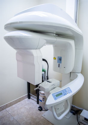 Cone beam Computed Tomography (CBCT) is one of the cutting edge diagnostics that are available at Oral and Maxillofacial Surgery of South Texas. Cone Beam CT is a medical imaging technique of producing a three-dimensional image of the internal structures of a solid object (such as the jaw and teeth). The X-rays are divergent, or opposing, and form a cone. For the assessment and planning of surgical implants, a Cone Beam scan can offer vital information for the surgeon to consider.
What is Dental Cone Beam CT?
Dental cone beam computed tomography (CT) is a special type of x-ray machine used in situations where regular dental or facial x-rays are not sufficient. This type of CT scanner uses a special type of technology to generate three dimensional (3-D) images of dental structures, soft tissues, nerve paths and bone in the craniofacial region in a single scan. Images obtained with Cone Beam CT allow for more precise treatment planning.
Cone beam CT provides detailed images of the bone and is performed to evaluate diseases of the jaw, dentition, bony structures of the face, nasal cavity and sinuses.
How should I prepare?
A Cone Beam CT examination requires no special preparation.
Prior to the examination, you may be asked to remove anything that may interfere with the imaging, including metal objects, such as jewelry, eyeglasses, hairpins, hearing aids, and any removable dental appliances.
Women should always inform their dentist or oral surgeon if there is any possibility that they are pregnant.
How does the procedure work?
During a cone beam CT examination, the C-arm or gantry rotates around the head in a complete 360-degree rotation while capturing multiple images from different angles that are reconstructed to create a single 3-D image.
You will not experience any pain during a cone beam CT exam, and you will be able to return to your normal activities once the exam is complete.
What are some common uses of the procedure?
- Assesment and planning for placement of dental implants.
- Surgical planning for impacted teeth.
- Diagnosing temporomandibular joint disorder (TMJ).
- Evaluation of the jaw, sinuses, nerve canals and nasal cavity.
- Detecting, measuring and treating jaw tumors.
- Determining bone structure and tooth orientation.
- Locating the origin of pain or pathology.
- Cephalometric analysis.
- Reconstructive surgery.
What are the benefits?
- The focused x-ray beam reduces scatter radiation, resulting in better image quality.
- A single scan produces a wide variety of views and angles that can be manipulated to provide a more complete evaluation.
- Cone beam CT scans provide more information that conventional dental x-ray, allowing for more precise treatment planning.
- CT scanning is painless, noninvasive and accurate.
- A major advantage of CT is its ability to image bone and soft tissue at the same time.
- No radiation remains in a patient’s body after a CT examination.
- X-rays used in CT scans should have no immediate side effects.